X-ray microtomography at INRAE Clermont-Ferrand
For more information, contact Eric BADEL (Eric.Badel@clermont.inrae.fr)
What is X-ray microtomography?
X-ray microtomography is a non-destructive method that allows access to the inner vision of an object (composition, arrangement, defects, porosity); that is to say without cutting the sample. The use of this technique is currently experiencing a boom, particularly in biology.
This imaging technique is based on the property of X-rays to pass through the material and be absorbed in function of the nature and density of the components they cross. A tomographic scan consists in the recording a series of digital radiographs of the sample at various angles. After a "reconstruction" computation, these data allow the 3D visualization, mapping real variation of attenuation of X-rays through the object.
| |
|
|
|
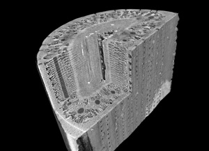
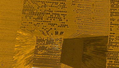
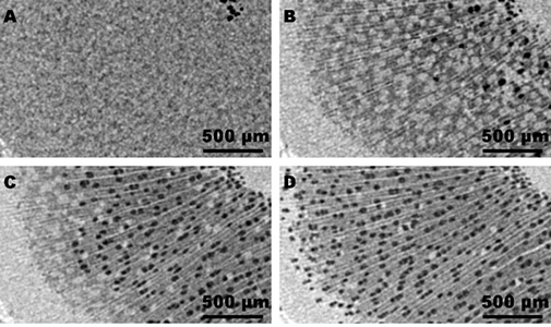
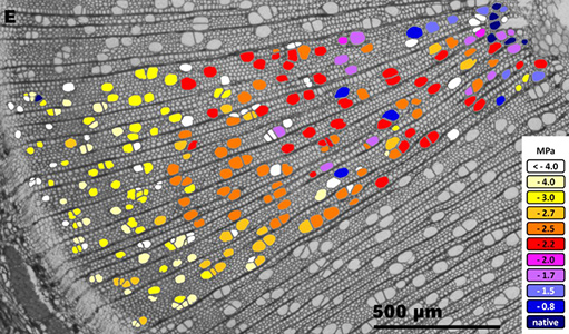
Structure of a pine needle Charra-Vaskou et al 2012 (Tree Physiology) | Distribution of summer embolism in an annual ring of Douglas wood Dalla-Salda et al 2014 (J Plant Hydraulics) |
Therefore, the internal structure of the object can be described qualitatively and quantitatively:
- Dimensional measurements in three dimensions
- Spatial distribution of the various phases of a heterogeneous material.
- Characterization of porosity (connectivity, etc)
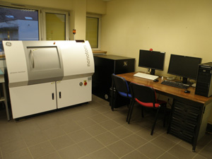
Microtomograph "Nanotom" installed at INRAE-Clermont-Ferrand
The characteristics of our Nanotom installed at INRAE in Clermont Ferrand can cover samples from sub-millimeter to ten centimeters with spatial resolutions ranging from 1 to 50 microns.
The device consists of a model microtomograph "Nanotom" Phoenix GE company and a set of six work stations for the acquisition and processing of the 2D and 3D images.
| |
|
|
|
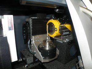
X-ray microtomography device at INRA Clermont-Ferrand | The sample room |
Some characteristics of microtomograph "Nanotom" installed at INRA-Clermont-Ferrand (Crouël site):
· An X-ray source-focus nano Phoenix GE (160kV, 15W, 0,9μm) equipped with interchangeable targets (tungsten or molybdenum) to pass through high density objects (metal, rocks, etc.) and for the adjustable scan of samples of low density as the original biological objects.
- An imaging (2000 x 2000 pixels) of Hamamatsu 50 microns spatial resolution. Ability to create a virtual imaging to increase the field of view.
- Reconstruction of the images on a cluster of four stations (32 GB RAM): 10003 voxels are reconstructed in few minutes
- Spatial resolution from 50 microns to 0,9μm.
- Maximum field of view: 80 mm.
- Scan time from a few minutes to one hour.
- Processing softwares and visualization of 2D and 3D images such as vgstudio, ImageJ, Matlab tools.
Examples of applications on the PHENOBOIS platform
The PIAF has developed a particular competence on the observation of biological objects and in particular on plants.
- Visualization of the architecture of the hydraulic network of a stem
| |
|
|
|
Grafted vine stem | 3D rendering of the hydraulic network architecture of a grafted vine stem |
- Visualisation de la propagation d’embolie à l’échelle cellulaire
| |
|
|
|
Monitoring of a poplar sample during a drought | Multispectral approach by combining X-ray microtomography with cytological sections Lemaire and al 2021 (Tree Physiol) |
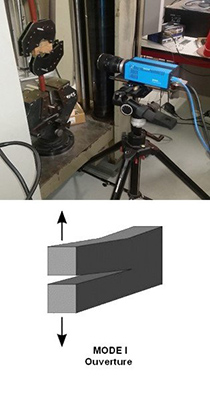
- 3D cracking monitoring and fracture toughness measurements
| | |
|
|
|
|
Mechanical fracture test | Transverse view of the crack | Longitudinal view of the crack |
- Characterization of biosourced materials
| |
|
|
|
Sample of composite made from corn fibers and a lignosulfate matrix | View of a cross section made in the 3D image obtained by microtomographic scanning. Tribot and al 2018 ( Industrial Crops and Products) |
- Archaeological objects: determination of the species (boxwood) and dating by dendrochronology of a spindle whorl
| |
|
|
|
Gallo-Roman spindle whorl (Bargoin Museum - Clermont-Ferrand) | Cross section of the spindle whorl made by X-ray microtomography |
- Archeology: Characterization of a 410 million year old fossil identified as the 1st tree on Earth (Armoricaphyton chateaupannense)
| | |
|
|
|
|
Fossil sample | 2D and 3D renderings | Modeling the conduction capacities of the hydraulic network elements of the stem Strullu and al 2014 (Botanical Journal of the Linnean Society) |
- Observation of various biological objects
| | |
|
|
|
|
Structure of a wheat ear by X-ray microtomography | Journey of a codling moth caterpillar in an apple tree | 3D rendering of the muscular system of Drosophila |
Phenobois :
https://www6.inrae.fr/phenobois
References :
Mantova M, Menezes-Silva PE, Badel E, Cochard H & Torres-Ruiz JM. 2021. The interplay of hydraulic failure and cell vitality explains tree capacity to recover from drought. Physiologia Plantarum 172, 247-257. ⟨hal-03109304⟩
Lemaire C. Quilichini Q., Brunel-Michac N., Santini J., Bertie L., Cartailler J., Conchon P., Badel E. and Herbette S. 2021. Plasticity of the xylem vulnerability to embolism in Populus tremula x alba relies on pit quantity properties rather than on pit structure. Tree Physiol. ⟨hal-03141908⟩
Ludovic Martin, Hervé Cochard, Stefan Mayr, Eric Badel. Using electrical resistivity tomography to detect wetwood and estimate moisture content in silver fir (Abies alba Mill.). Annals of Forest Science, Springer Nature (since 2011)/EDP Science (until 2010), 2021, 78 (3), pp.1-17. ⟨hal-03318957⟩
Billon LM, Blackman CJ, Cochard H, Badel E, Hitmi A, Cartailler J, Souchal R, Torres-Ruiz JM. 2020. The DroughtBox: a new tool for phenotyping residual branch conductance and its temperature dependence during drought. Plant, Cell and Environment 43: 1584– 1594 ⟨hal-02625372⟩
Myriam Moreno, Guillaume Simioni, Maxime Cailleret, Julien Ruffault, Eric Badel, et al.. Consistently lower sap velocity and growth over nine years of rainfall exclusion in a Mediterranean mixed pine-oak forest. Agricultural and Forest Meteorology, 2021, 308-309, pp.108472. ⟨10.1016/j.agrformet.2021.108472⟩. ⟨hal-03321323⟩
Sergent A.S., Varela S., Badel E., Barigah T., Cochard H., Dalla-Salda G., Delzon S., Fernández M.E., Guillemot J., Gyenge J., Lamarque L.J., Meier A.M., Rozenberg P., Torres-Ruiz J.M., Martin N. 2020. A comparison of five methods to assess embolism resistance in trees. Forest Ecology and Management. 468. ⟨hal-02769656⟩
Francois Courbet, Nicolas Martin, Guillaume Simioni, Claude Doussan, Jean Ladier, et al.. Agir sur la sensibilité à la sécheresse par la sylviculture. Forêt Entreprise, Forêt Privée Française, 2019, 249 (Novembre - décembre), pp.46-48. ⟨hal-02394661⟩
Muries Bosch B., Mom R., Benoit P., Brunel-Michac N., Cochard H., Roeckel-Drevet P., Petel G., Badel E., Fumanal B., Gousset A., Julien J.L.,, Label P., Auguin D., Venisse J.S. 2019. Aquaporins and water control in drought-stressed poplar leaves: A glimpse into the ex-traxylem vascular territories. Enviv. and Exp Bot, 162, pp.25-37. ⟨hal-02168616⟩
Mayr S., Schmid P., Beikircher B., Feng F., Badel E. 2019. Die hard: timberline conifers survive annual winter embolism. New Phytol 226(1):13-20. ⟨hal-02394658⟩
Tribot A., Delattre C., Badel E., Dussap C.G., Michaud P., de Baynast H. 2018 Design of experiments for bio-based composites with lignosulfonates matrix and corn cob fibers. Industrial Crops and Products.123: 539-545. ⟨hal-01915473⟩
López R., Nolf M., Duursma R., Badel E., Flavel R.J., Cochard H., Choat B. 2018 Mitigating the open vessel artefact in centrifuge based measurement of embolism resistance. Tree Physiol 39(1):143-155. ⟨hal-01854603⟩
Lamarque L., Corso D., Torres-Ruiz J.M., Badel E., Brodribb T., Burlett R., Charrier G., Choat B., Cochard H., Gambetta G., Jansen S., King A., Lenoir N., Martin-StPaul N., Steppe K., Van den Bulcke J., Zhang Y. and Delzon S. 2018 An inconvenient truth about xylem resistance to embolism in the model species for refilling Laurus nobilis L. Annals of Forest Sciences. 75-88. ⟨hal-02288736⟩
Torres-Ruiz J.M., Cochard H., Choat B., Jansen S., Tomášková I., Padilla-Díaz C.M., Badel E., Burlett R., King A., Lenoir N., Martin-StPaul N.K. and Delzon S. 2017. Xylem resistance to embolism: presenting a simple diagnostic test for the open vessel artefact. New Phytol. 215:484-499. ⟨hal-02288736⟩
Torres-Ruiz J.M., Cochard H., Fonseca E., Badel E., Gazarini L. and Vaz M. 2017. Differences in functional and anatomical traits within co-occurring Cistus species to face drought stress. Tree Physiol. 37(6):755-766. ⟨hal-01606013⟩
Torres-Ruiz J.M., Cochard H., Mencuccini M., Delzon S., Badel E. 2016. Direct observations and modelling of embolism spread between xylem conduits: a case study in Scot pine. PCE. 39(12):2774-2785. doi:10.1111/pce.12840 ⟨hal-01512094⟩
David-Schwartz R., Paudel I., Delzon S., Cochard H., Mizrachi M., Zemach H., Lukyanov V., Badel E., Capdeville G., and Cohen S., 2016. Indirect evidence for genetic differentiation in vulnerability to embolism in Pinus halepensis. Frontiers in Plant Sciences. 7, 13 p. ⟨hal-01512018⟩
Hochberg U., Herrera J.C., Cochard H. and Badel E. 2016. Short-time xylem relaxation results in reliable quantification of embolism in grapevine petioles and sheds new light on their hydraulic strategy. Tree Physiol. 36 (6), pp.748-55. ⟨hal-01343636⟩
Choat B., Badel E., Burlett R., Delzon S., Cochard H., Jansen S., 2016. Non-invasive measurement of vulnerability to drought induced embolism by X-ray microtomography. Plant Physiol. 170(1):273-282. ⟨hal-02637984⟩
Hervé H. Cochard, Sylvain S. Delzon, Eric Badel. 2015. X-ray microtomography (micro-CT): a reference technology for high-resolution quantification of xylem embolism in trees. Plant, Cell and Environment, Wiley, 38 (1), pp.201-206. ⟨hal-02635290⟩
Charra-Vaskou K., Badel E., Charrier G, Ponomarenko A., Bonhomme M., Foucat L., Mayr S., Améglio T. 2015. Cavitation and water fluxes driven by ice water potential in Juglans regia during freeze-thaw cycles. JXB.67(3),739-750. ⟨hal-01261003⟩
Dalla-Salda G., Fernández M.E., Sergent A.S., Rozenberg P., Badel E. and Martinez-Meier A. 2014. Dynamics of cavitation in a Douglas-fir tree-ring: transition-wood, the lord of the ring? Journal of Plant Hydraulics 1: e-0005. ⟨hal-01095363⟩
Torres-Ruiz J.M., Cochard H., Mayr S., Beikircher B., Diaz-Espejo A., Rodriguez-Dominguez C.M., Badel E., Fernández J.E, 2014. Vulnerability to cavitation in Olea europaea current-year shoots: more support to the open-vessel artefact with centrifuge and air-injection techniques. Physiologia Plantarum 152 : 465-474. ⟨hal-01123401⟩
Strullu-Derrien C., Kenrick P., Tafforeau P., Cochard H., Bonnemain J.L., Le Hérissé A., Lardeux H. and Badel E., 2014. The earliest fossil wood and its hydraulic properties documented in a 407 million year old plant from the Armorican Massif, France. Botanical Journal of the Linnean Society 175, 423–437. ⟨insu-01018347⟩
Cochard H., Badel E., Herbette S., Delzon S., Choat B., Jansen S. 2013. Methods for measuring plant vulnerability to cavitation: a critical review. J Exp Bot. 64 (15): 4779–4791. ⟨hal-00964655⟩
Charra-Vaskou K., Badel E., Burlett R., Cochard H., Delzon D., Mayr S., 2012. Hydraulic efficiency and safety of vascular and non-vascular components in Pinus pinaster leaves. Tree Physiol, 32 (9): 1161-1170. ⟨hal-00964504⟩
Copyright © 2012, INRA, All rights reserved


